

Osteochondral Injury of the Ankle Joint Symptoms & Treatment
An osteochondral injury happens when the smooth cartilage that lines a joint and the bone beneath it are damaged. In the ankle, this often follows a twist, fall, or severe sprain that harms the cartilage surface. The injury may lead to pain, swelling, stiffness, and trouble walking. Many mild osteochondral lesions improve with rest, ice, bracing, and physical therapy, while more severe cases may need surgical care.
Start with our quick Symptom Assessment or connect directly with an Upswing Coach today.
Request an AppointmentReady to take the next step?
Start your symptom assessment or connect with a coach instantly.
Find Relief TodayOverview
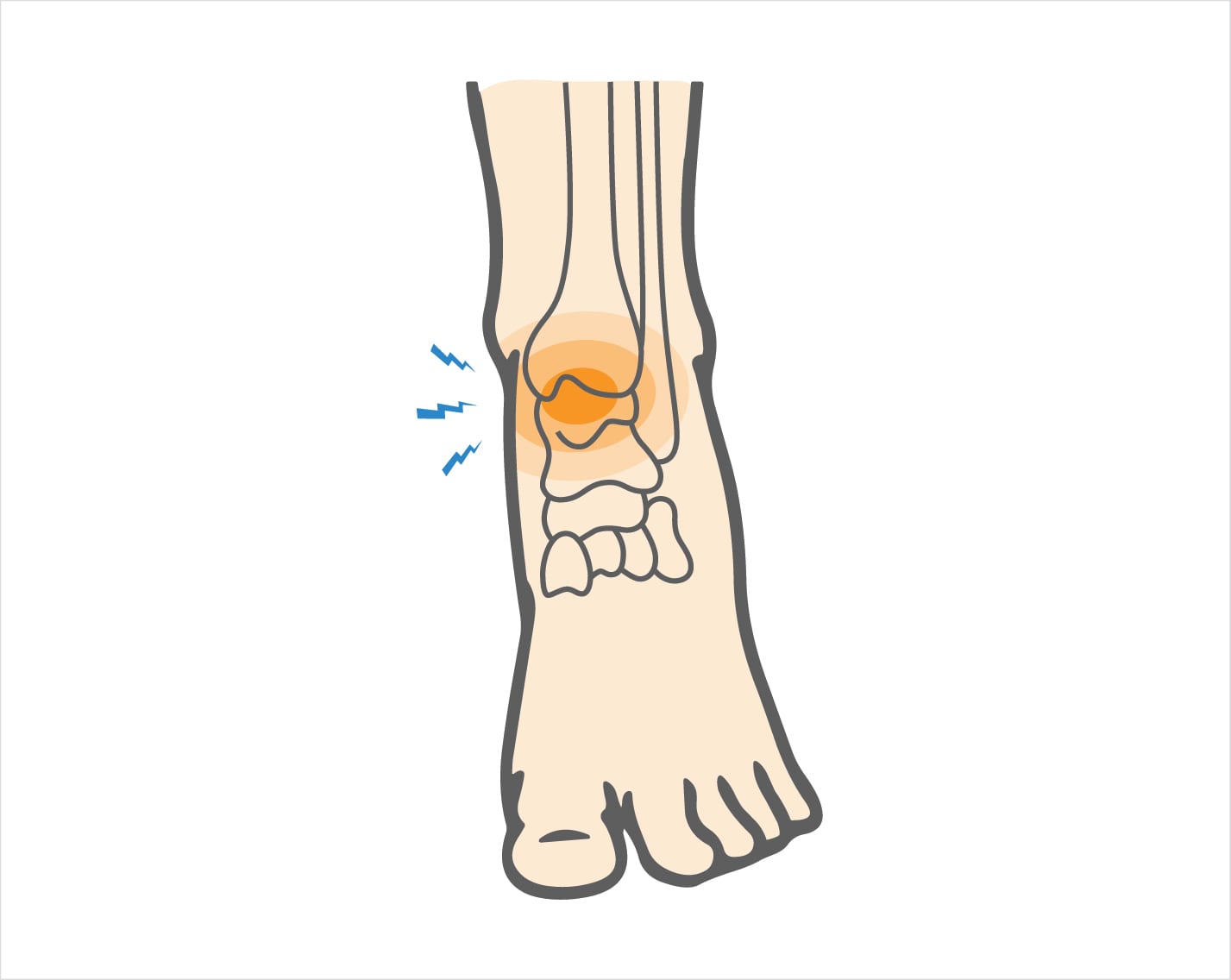
An osteochondral injury is damage to both the cartilage surface of a joint and the layer of bone beneath it. In the ankle, this occurs on the top of the talus, a small bone that connects your leg to your foot,, where smooth cartilage allows the joint to move without strain. When this cartilage or bone is injured, it forms what is known as an osteochondral lesion of the ankle.
These injuries may follow a sudden impact, a severe ankle sprain, or a fracture. In some people, chronic ankle instability from repeated sprains gradually wears down the cartilage. Osteochondral damage can range from mild cartilage softening to deep lesions in which cartilage and bone fragments separate within the joint. If a fragment breaks free, it may cause locking or catching in the ankle and limit movement.
Although most common in the ankle, similar damage can also occur in the knee, where it is referred to as an osteochondral lesion of the knee.
What causes Osteochondral Injury of the Ankle Joint?
Most osteochondral injuries develop after a twisting motion or sudden impact that produces an ankle sprain. When the joint is forced out of position, the talus may strike the tibia or fibula. This contact can bruise or tear the smooth articular cartilage and may create a small fracture through the cartilage and the bone beneath it.
How the Injury Occurs
- A forceful twist or roll of the ankle
- Impact during sports
- Landing awkwardly from a jump
- Direct trauma to the joint
Common Everyday Causes
- Slipping on stairs
- Losing balance while walking on uneven ground
- Chronic ankle instability from previous sprains
- Wearing improper footwear with poor ankle support
Sports Where Osteochondral Injuries Are Most Common
Soccer – Quick direction changes and ankle-to-ankle contact often lead to twisting injuries.
Football – High-impact tackles and sudden pivots can damage the cartilage surface.
Rugby – Repeated collisions and unstable footing increase the risk of osteo lesions.
Basketball – Jumping, landing, and rapid lateral movement frequently strain the ankle joint.
Symptoms
Early osteochondral injuries often feel similar to an ankle sprain, with a dull ache that becomes worse when you put weight on the joint. The symptoms can vary depending on how much cartilage and bone have been damaged.
Common symptoms include:
- Persistent ankle pain
- Swelling that does not fully go away
- Limited range of motion or stiffness
- Difficulty walking or standing for long periods
- A clicking, catching, or “locking” sensation in the ankle
When cartilage fragments become loose, the ankle may feel unstable or may get stuck in a certain position.
When to see a doctor
You should see a doctor if your ankle remains swollen, painful, or difficult to walk on after an injury. During the visit, the doctor will ask how the injury occurred, what activities you take part in, and whether you have had ankle problems in the past.
The examination may include gentle pressure on the joint to check for tenderness or ligament injury. Your doctor may also order imaging tests:
- Weight-bearing X-rays to look for narrowing of the joint space or small defects in the bone
- MRI to identify cartilage damage, bone swelling, and the full extent of the osteochondral injury
These studies help determine how severe the lesion is and guide the plan for treatment.
Non-operative treatment
Most mild osteochondral injuries can be managed without surgery. Early care aims to reduce pain and protect the ankle while the cartilage and bone begin to heal.
Common non-surgical treatments include:
- Ice: Apply for 20 minutes every 2–3 hours
- Elevation: Raise the ankle to reduce swelling
- Compression: Use an Ace bandage or ankle wrap
- NSAIDs: Medications such as ibuprofen or naproxen to ease pain
- Bracing or casting: To stabilize the ankle and prevent further injury
- Physical therapy: To restore strength, balance, and range of motion
A gradual return to weight-bearing is important, starting slowly and avoiding activities that cause pain.
Try these exercises to help address your condition:
Below is a PDF of the Exercise Program
Surgical Treatment
For more serious osteochondral lesions of the ankle, your doctor may refer you to an orthopedic surgeon. Surgery is often considered when loose bone or cartilage fragments are present or when the joint surface has a deep defect.
Common surgical options include:
- Debridement: Removing loose fragments of cartilage or bone
- Microfracture or drilling: Creating small holes in the bone to stimulate new cartilage growth
- Osteochondral grafting: Transplanting healthy bone and cartilage to fill the lesion
Your surgeon will determine the most suitable procedure based on the size and location of the osteo lesion.
Recovery
Recovery time depends on how severe the injury is and whether surgery was needed. Mild lesions may heal within a few weeks, while more significant injuries can require several months. Most rehabilitation plans include:
- Protecting the ankle with crutches or a boot
- Gradually returning to weight-bearing activities
- Stretching and strengthening exercises
- Balance and stability training through physical therapy
A careful, steady recovery helps protect the healing cartilage.
Dr. Jay Kimmel is a board-certified orthopedic surgeon specializing in sports medicine, arthroscopic surgery, and shoulder and knee disorders. He completed his orthopedic training at New York-Presbyterian/Columbia University Medical Center and a Sports Medicine Fellowship at Temple University.
Dr. Kimmel previously served as the Director of the Connecticut Sports Medicine Institute at Saint Francis Hospital and has held faculty appointments as Clinical Assistant Professor in the Departments of Orthopedics and Family Medicine at the University of Connecticut. He has extensive experience caring for athletes as a team physician for high school and collegiate programs and continues to teach in the athletic training departments at Westfield State University and Springfield College.
Find the Support You Need — Right When You Need It
Whether you’re managing pain for the first time, need ongoing guidance, or require expert medical care, we’re here to help you every step of the way.
ORTHO DIRECT
Video visit with an orthopedic doctor for advice and a care plan.
$30
/MonthMRI DIRECT
Fast, affordable MRI with orthopedic review. No insurance required.
$499
/MonthFrequently Asked Questions
How do I know if my ankle pain is from an osteochondral lesion?
Persistent swelling, pain that doesn’t improve after a sprain, stiffness, or ankle locking may indicate an osteochondral ankle lesion.
Can an osteochondral lesion heal on its own?
Smaller lesions may heal with rest, bracing, and physical therapy, while larger or displaced lesions often require surgical treatment.
How do doctors diagnose an osteochondral injury?
Doctors use X-rays and MRI scans to identify cartilage damage, bone swelling, fragment displacement, and the severity of the osteo lesion.
What are the treatment options for osteochondral injuries?
Treatment ranges from rest, ice, and physical therapy to surgical procedures such as debridement, microfracture, or cartilage grafting.